October 2017, Vol. 244, No. 10
Features
Real-Time Imaging of Methane Gas Leaks Using a Single-Pixel Camera

The ability to image invisible gases has applications in industrial and environmental monitoring settings, but is technologically challenging to embed in a low-cost device. For example, imaging methane gas has applications among gas utility companies for routine pipeline monitoring and storage facility inspection.
Video rate gas imaging conveys the direction of dispersal and hence the location of a leak source, helping users to improve their efficiency of response to hazardous events. Conventional approaches to detecting methane gas leaks have mainly been based upon flame ionization detectors (FIDs), but such technology measures concentration at only a single point, making locating the source of the leak a difficult and slow process.
One approach to gas imaging is to use a focal plane array (FPA) to image the methane directly. These array-based systems can be passive, based on the absorption of ambient background radiation or active, based on the optical absorption of laser light used as an illumination source. Passive systems for imaging methane leaks have been demonstrated using infrared cameras employing outdoor thermal background radiation.
However, the sensitivity of such systems is particularly susceptible to variations in this background temperature, which at certain times during the daily cycle may significantly reduce the sensitivity of the instrument. In an active system, the use of InGaAs laser diodes, emitting at 1.6 µm, are a convenient option for the illumination source due to their compact size, allowing the development of portable systems which can detect methane in the short-wave infrared (SWIR) spectrum. FPAs that operate at SWIR wavelengths are less well-developed than their visible wavelength equivalents, having a much-reduced resolution and increased cost.
As an alternative to using a FPA, it is possible to use a single photo-detector and an infrared laser, wavelength tuned to an absorption line of the gas, which is raster scanned over a scene and the resulting back-scattered light collected and measured. Infrared laser light suitable for detecting methane can be provided by many different sources including, compact distributed feedback InGaAs laser diodes operating at wavelengths around 1.6µm, optical parametric oscillators operating at wavelengths around 3.4µm or carbon dioxide lasers operating around 10µm. Of these sources the laser diode provides a cost-effective option.
Rather than raster scanning using galvo optics, the techniques of single-pixel/computational imaging combine pattern projection and single-pixel detectors to reconstruct images. One approach is to use a sequence of masks to structure the illumination light which is then projected onto the scene and to measure the resulting backscattered light using a single photodetector.
The single detector, or single-pixel, is used to measure the level of correlation between a set of patterns and the scene. Knowledge of these patterns and the corresponding correlation can be computationally inverted to enable the reconstruction of the image of the scene. Here, we define our masks [based on Hadamard matrices], which are binary functions, the masks from which form a complete orthonormal set and where each pattern contains an equal number of +1’s and −1’s, representing ‘on’ and ‘off’ respectively, for each mask.
Single-pixel imaging has the benefit of being able to image at wavelengths where multi-pixel image sensors are unavailable or costly, such as in the terahertz band or SWIR. The availability and low cost of single-pixel detectors operating in the SWIR spectral region make single-pixel cameras much more promising as a gas-imaging device.
Here, we use a digital micro-mirror device (DMD) to pattern the output of an infrared diode laser, tuned to a methane absorption line at 1.651 µm, which is then projected onto a scene containing various sources of methane. An amplified InGaAs photodiode is used as the single-pixel to measure the laser light backscattered from the scene, some of which passes through any sources of gas present.
DMDs, consisting of hundreds of thousands of individually addressable moving micro-mirrors, were originally developed for the display industry but have also found applications in other areas including wavelength multiplexing, real-time infrared imaging, multi-object spectroscopy and also applicable to astronomical observations. They offer a method of modulating light that is fast and works over a broad range of wavelengths.
DMDs can be used to implement masks representing +1 (transmitted light) and 0 (blocked), but not +1 and −1 required for a Hadamard set. To overcome this, each mask is displayed as a pair of patterns where each pattern is followed by its negative, allowing a differential signal to be obtained.
The number of mask patterns required to fully reconstruct an image frame is proportional to the linear resolution squared (this number is doubled for the differential case) and hence, limits the frame rate for high-resolution reconstructions. However, DMDs are commercially available, having display rates of about 22 kHz, which for relatively low-resolution applications allows near-video rate image reconstruction on a standard performance computer.
For the case of locating gas leaks, the resolution of the image is of secondary importance. More important is the frame rate of the system which can image quickly enough to locate the source of the leak before the gas has time to diffuse away.
We demonstrate our single-pixel imaging system in the laboratory at two different reconstruction resolutions. We image a static scene containing glass cells filled with known concentrations of methane in air, at a resolution of 64 by 64, which shows the individual cells and their contents.
We demonstrate real-time imaging of a gas leak from a ¼-inch diameter tube of about 0.2 liters/minute, at a resolution of 16 by 16 (512 masks corresponding to a full set of 16 by 16 pixel patterns + inverse). Although low-resolution image reconstructions are sufficient for determining the source of a gas leak, it is desirable to combine this information with high-resolution color images of the scene. For this reason, a visible camera is used to provide an image for the operator, upon which the gas data is overlaid.
Experimental Configuration
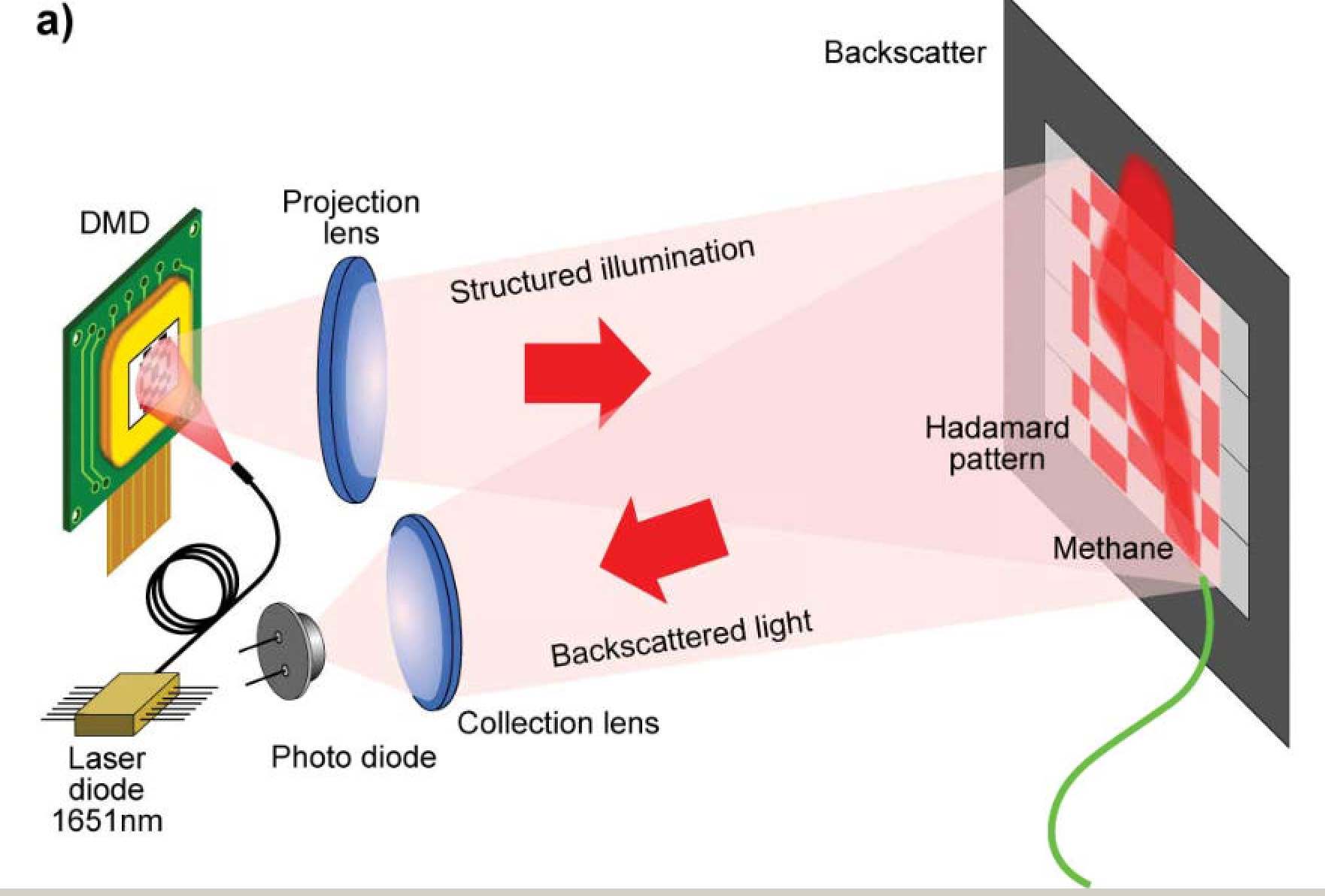
Figure 1(a) shows an illustration of the single-pixel gas imaging camera. An infrared source consisting of a fiber coupled InGaAs laser diode (LASER 2000, LAS-022527) is used to illuminate a high-speed DMD (Vialux, V-7000). The wavelength of the laser can be coarsely tuned with temperature and finely tuned with current, both controlled using the laser driver (Thorlabs, CLD1015).
The wavelength is initially set to correspond to a methane absorption transition at 1.651 µm, and applying a square wave signal of ± 0.014 V to the laser diode drive current modulation input allows the laser to be tuned on and off of the methane absorption, allowing a differential gas measurement. The tuning rate of the laser is about 12 pm/mA and the 0.014 V modulation corresponds to a wavelength shift of 0.05 nm, larger than the absorption line width of methane at atmospheric pressure. (Fig. 1)

Figure 1: a) Schematic of optical configuration, b) An optical head containing the DMD, laser fiber, CMOS camera and single-pixel detector.
Rather than raster scanning the laser over the scene, we structure the illumination using patterns displayed on the DMD. A singlet lens, diameter 30mm and 50mm focal length, with an anti-reflection (AR) coating for infrared, is used to project the patterns onto the scene. The sequence of patterns is preloaded onto the memory buffer of the DMD and then displayed at 20 kHz. The DMD chip resolution is 1,024 by 768 with a mirror pitch = 13.7µm (DLP, Discovery 4100). The mirrors are binned according to the desired reconstructed resolution, using a 768 –by-768 area of the chip. An AR coating on the window of the DMD chip reduces losses of the 1.65µm light.
A simple low-cost, uncooled, amplified InGaAs photodiode (spectral response 800 nm–1800 nm) (Thorlabs, PDA20CS-EC) is used to measure the total intensity of the back-scattered laser light from the scene, collected using a singlet lens, diameter 30mm and 30mm focal length, having a similar AR coating to the projection lens. Signals from the photodiode are read using a 16-bit data acquisition module (DAQ) (National Instruments, NI USB-6366), capable of sampling at 2.0 MS/s.
The signal capture is synchronized to the pattern update on the DMD. The same DAQ is also used to provide the square wave signal for the laser drive current modulation, enabling the laser to be tuned on and off of the methane absorption line.
The gas-imaging camera is packaged as a custom 3-D printed unit containing the DMD, laser fiber mount, projection lens and single-pixel backscatter detector (Figure 1 (b)). The unit also includes a high-resolution color CMOS camera to provide a navigation/guide image. The gas image information is aligned and overlaid on the images from the color CMOS camera (JAI GO-5000C-USB). Custom LabVIEW (National Instruments) interface software controls the DMD, collects all single-pixel intensity measurements from the DAQ, reconstructs the gas image and overlays an up-sampled smoothed gas image on the color image of the scene obtained from the CMOS camera.
Results
We constructed a scene consisting of four glass sample cells containing known mixtures of methane in air (0%, 25%, 50% and 100% volume), all at atmospheric pressure. The cells were all of 20-mm diameter, 10-mm thick and arranged in front of a background, which consisted of a black cardboard screen with strips of infrared reflecting tape covering the central region.
The size of the infrared reflecting region was 250 mm by 200 mm and the background was located 1 meter from the camera lens. Also included within the scene were two ¼-inch diameter PTFE tubes used to deliver gas leaks of a predetermined flow rate (the two gases used were pure methane and nitrogen). Needle valves with floating ball indicators allowed the flow rates through these tubes to be controlled over the range 0.1-5.0 liters/min. Figure 2 (a) shows a photo of the gas cell arrangement in front of the reflective background. Figure 2 (b) shows the methane absorption spectra at around 1.65 µm, obtained from the HITRAN database. (Fig. 2)
The DMD memory buffer was initially loaded with 8,192 patterns, corresponding to a 4,096 Hadamard set at a resolution of 64 by 64 and their inverse patterns. At this resolution, all four sample cells are clearly visible (Figure 2 (c) and Figure 2 (d)). For every alternate reconstruction frame, the wavelength of the laser was tuned either on or off the methane absorption using the laser diode current modulation input on the controller, reconstructed for an 8,192 pattern sequence. By taking the difference of the single-pixel intensity measurements between alternate frames, images can be obtained of the gas only as shown in Figure 2 (e).
The three sample cells containing methane are visible in the reconstructed difference gas image. The 64-by-64 resolution images are then averaged over five frames, smoothed and up-sampled as shown in Figure 2 (f). The 5-by-5 kernel used to smooth the gas image is shown in the inset of Figure 2 (f).

Figure 2: a) Gas sample cells located in front of the infrared reflective background; b) methane absorption data at 1.65µm, obtained using the HITRAN database; c-d) on- and-off-absorption infrared images of gas sample cells; e) gas image at 64 by 64 resolution; f) smoothed, averaged and up-sampled gas image. Inset shows the 5 by 5 smoothing kernel used.
In many imaging applications a high-resolution is the preferred option. However, in the case of single-pixel imaging, there is a trade-off between the resolution and frame rate of the reconstructed images, as the number of mask patterns required is proportional to the square of the linear resolution.
In addition, the signal-to-noise ratio (SNR) scales in inverse proportion to the linear resolution for constant pattern, display time. Both of these factors mean that lower resolutions are a viable and attractive option for single-pixel imaging. Figure 3 shows a single frame from the gas-imaging camera.
Figure 3 (a) shows the low-resolution (16-by-16) gas image, Figure 3 (b) shows the smoothed gas image and Figure 3 (c) shows the gas image overlay. Here, a total of 512 patterns are loaded onto the DMD, resulting in a reconstruction frame rate of 25 Hz. The differential images are again averaged over five frames and are smoothed using a 3-by-3 kernel as shown in inset of Figure 3 (b). Again, all of the glass cells containing methane are clearly visible, which allows the gas images to be correctly scaled and aligned with high-resolution images from the color camera.
To help the operator in identifying the gas sources, the up-sampled images are thresholded and color-coded red before being overlaid on the color image, as shown in Figure 3 (c). The CMOS camera covers a slightly larger field-of-view compared to the single-pixel camera, which allows a larger area of the scene to be viewed. The green outline box overlaid on the high-resolution image in Figure 3 (c) indicates the active area over which the gas can be detected. (Fig. 3)

Figure 3: a) Low-resolution (16-by-6) reconstructed image of gas cells and gas leak; b) smoothed, averaged and up-scaled gas image (512-by-512), smoothing kernel shown in figure inset; c) color-coded gas data overlaid onto a high-resolution color image from a CMOS camera.
In addition to imaging sample cells of fixed methane concentration, we simulate a gas leak by introducing gas from a pressurized cylinder containing 100% methane, delivered to the scene via a length of ¼-inch diameter PTFE tube. Again the images are averaged, smoothed and up-sampled before being thresholded and overlaid on the high-resolution color image.
To demonstrate that our imaging system is sensitive to methane only, we introduce a second gas leak to the scene from a pressurized nitrogen cylinder and show that only the methane is visible in the reconstructed image. Figure 4(a) shows representative frames from a video of a gas leak for a methane flow rate of 0.2 liters/min (delivered via the green tube shown on the right-hand side of the frames). The reconstruction and smoothing frame rate for a 16-by-16 resolution is 25Hz and allows the source of the leak to be easily identified.
Nitrogen is introduced to the scene via the red tube, shown on the left-hand side of the frames, and is fixed at a flow rate of 2 liters/min. While the methane leak is clearly visible, the nitrogen leak is completely undetected, even when both leaks are simultaneous, as we are sensitive only to the methane absorption rather than the temperature difference between the escaping gas and the background.
The effect of the nitrogen leak on the methane can be seen in later frames of Figure 4(a) in which the gas plume is seen to blow toward the right. The frame rate of our system is sufficient that we are relatively insensitive to motion blur from a dynamically changing scene. As a demonstration, we image a dynamic scene where one of the gas sample cells (containing 100% methane) is moved by hand across the field of view. Figure 4(b) shows selected frames from a recorded movie where only the gas cell is imaged despite other parts of the scene changing in real-time. (Fig. 4)

Figure 4: a) Frames from a low-resolution (16-by-16) computational image of a gas leak, overlaid onto a high-resolution color image from a CMOS camera; b) frames from imaging a dynamic scene where a gas sample cell is moved across the field of view.
Conclusions
We have demonstrated that a single-pixel imaging system can be used to image gas leaks in real-time. Reducing the reconstructed resolution of the gas images to 16×16 increases the frame rate, allowing the gas to be imaged in a dynamic scene. Smoothing the 16-by-16 gas data still gives images of sufficient quality to allow the user to identify the source of the leak.
An alternative method for increasing the frame rate while maintaining a higher resolution is to use compressive sensing techniques, which exploit knowledge of the basis in which an image can be sparsely represented. A reduction in the numbers of mask patterns can also be achieved using adaptive sampling, where pattern choice is based upon previous measurements.
A limitation of single-pixel imaging is the reduction in the SNR of the reconstructed images as the resolution is increased. Micro-scanning sets of lower resolution patterns enables an improvement in SNR with only a moderate sacrifice in reconstructed resolution. This provides a way to optimize the trade-off between SNR and resolution to suit the conditions of the scene.
Various research groups have investigated methods to enhance the performance of FPAs for infrared imaging. A SWIR detector having an increased sensitivity and angle-of-view, in addition to a higher power laser for the illumination, should allow imaging of gas over longer distances, over larger areas and with an uncooperative backscatter. In addition, the use of a widely tunable infrared laser source will allow the imaging of various gases, thus extending the range of possible applications.
Originally published by Optics Express, Vol. 25, Issue 4, pp. 2998-3005 (2017) https://doi.org/10.1364/OE.25.002998


Comments